Dermoscopy opens up a new dimension on clinical morphology of skin lesions. Digital follow-up examinations, computer-aided diagnosis, and teledermoscopy are new facilities that will change the current management of skin cancer…
A good argument can be made for skin cancer over diagnosis. And, according to the studies linked and excerpted below, a good argument can be made for the importance of dermoscopy for a skin cancer diagnosis.
Your concern is simply to insure an accurate skin cancer diagnosis regardless. If that mole on your shoulder or face was checked out by your doctor and he/she gave you the all clear, you want to make sure that he/she is correct. By the same token, if your doctor told you that your mole was skin cancer, you want to make sure he/she is correct.
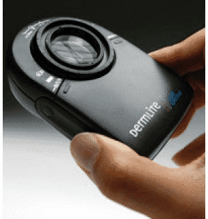
I had an appointment with a dermatologist last week. An intern came into the room before the dermatologist. Dr. Kamel pulled out his dermoscope and studied ever mark I had a concern about. And several lesions that I didn’t see.
When the dermatologist entered the room he and Dr. Kamel talked and studied my skin top to bottom. The dermatologist recommended that three spots on my head be frozen (cryotherapy) and that three suspicious looking moles be removed and biopsied.
The bottom line is that I am fine. For now. But the dermascope was an integral part of my skin examination.
Non-Melanoma Skin Cancer at a Glance-
- Risks– UV Exposure, HPV, Genetics, Skin Pigment, Immunosuppression, Radiation Therapy, Age, Previous Skin Cancer,
- Symptoms– Itching, Bleeding, Shape (A,B,C,D,E).
- Diagnosis– Visual inspection (A,B,C,D,E), Skin Biopsy (Shave, Punch, Incisional/Excisional)
- Prognosis- Staging-
- Therapy– Conventional, Non-Conventional, Int, Alternative
The good news in either scenario above you have choices. There are a host of evidence-based, non-toxic therapies to reduce your risk of non-melanoma skin cancer becoming melanoma. Further, many of these non-toxic therapies have been shown to heal sun damaged skin.
I am a survivor of a completely different cancer. Many of the therapies I underwent for this cancer increased my risks for melanoma. I had a mole removed from my face a few years ago.
You can believe I take my skin cancer risk seriously. I take many evidence-based, non-toxic therapies to reduce my future risk of melanoma.
To Learn More About Actinic Keratosis- click now
Have you been diagnosed with non-melanoma or melanoma skin cancer? Scroll down the page, post a question or comment and I will reply to you ASAP.
Thanks,
David Emerson
- Cancer Survivor
- Cancer Coach
- Director PeopleBeatingCancer
Recommended Reading:
“A dermatoscope is a hand-held visual aid device a doctor or person can use to examine and diagnose skin lesions and diseases, such as melanoma. It can also help a person examine the scalp, hair, and nails…
Examination
Dermatoscopes use light and magnification to help a dermatologist see how a person’s skin looks in more detail.
Dermatoscopes help show details in the outer layer of skin that would not be visible to the naked eye.
Diagnosis
Since dermatoscopes can enhance a doctor’s view of the skin, they can aid in the diagnosis of skin conditions, such as melanoma.
In one 2018 review, researchers found that using a dermatoscope was more effective in diagnosing melanoma than a simple visual inspection of a skin lesion.
One 2019 review found that a dermoscopy, a method that uses a dermatoscope, can be effective in diagnosing cancerous and noncancerous skin lesions.
How does a dermatoscope work?
A dermatoscope is a hand-held device. It features a light source and a magnifier and works a little like a magnifying glass.
Magnifier
According to a 2015 studyTrusted Source, a traditional dermatoscope magnifies the view of the skin by 10 times. Video dermatoscopes can increase this to around 70–100 times.
Light
The light on the dermatoscope helps illuminate the skin in a special way, allowing a doctor to examine lesions without the light bouncing off of dry or oily skin, which would make it more difficult to see.
Pictures
Dermatoscopes can also take pictures as people use them. Doctors may examine these photos later.
Lemonaid has a team of licensed medical professionals who can prescribe name brand or generic ED medication. Get 50% off your first shipment, plus fast, free & discreet delivery right to your door.
What happens during a dermatoscopy?
When a doctor uses a dermatoscope to examine a person’s skin, the procedure is called a dermatoscopy.
First, a doctor will apply some gel, alcohol, or water to a person’s skin.
Next, a doctor will typically turn on the light on the device and hold the device’s lens over the area of skin they are examining. It will not hurt.
Sometimes, it will be necessaryTrusted Source for the doctor to gently touch the skin with the dermatoscope.
The device will provide light and magnification so the doctor can examine areas of interest or concern on the skin, such as a lesion. A doctor may also take photos of the area.
“The purpose of this review is to highlight recent advances in dermoscopy, reviewing primary research articles published in the last year…
Dermoscopy opens up a new dimension on clinical morphology of skin lesions. Digital follow-up examinations, computer-aided diagnosis, and teledermoscopy are new facilities that will change the current management of skin cancers in general and melanoma in particular. Dermoscopy in the hands of experienced physicians has higher discriminatory power than naked-eye examination to detect skin cancers.”
“Dermoscopy could assist osteopathic primary care physicians improve their diagnostic accuracy for skin cancer, thus preventing unwarranted referrals to specialists; however, more knowledge to encourage the use of dermascopes is warranted…
Survey data indicated 54% (n = 410) of participants had heard of a dermascope, while 15% (n = 123) had used one in the past and 6% (n = 193) were currently using one in practice. Forty-eight percent of participants (n = 360) reported seeing at least 10 patients monthly for skin lesions suspicious of skin cancer and 53% (n = 402) reported being confident or very confident in differentiating between cancerous and noncancerous skin lesions…
Morris and colleagues found that graduating from medical school after 1989 and having more confidence in differentiating skin lesions were statistically significant multivariate predictors for dermascope use…


