fbpx

Diagnosed with Cancer? Your two greatest challenges are understanding cancer and understanding possible side effects from chemo and radiation.  Knowledge is Power!

Learn about conventional, complementary, and integrative therapies.

Dealing with treatment side effects? Learn about evidence-based therapies to alleviate your symptoms.

Click the orange button to the right to learn more.

When Actinic Keratosis (Pre-Skin Cancer) is Not Actinic Keratosis

Share Button

Got Spots (Actinic Keratosis-pre-skin cancer) on Your Bald Head? Get a Closer Look to Distinguish AK vs. IC vs. SCC-

Image showing an actinic keratosis

I’m bald. I have spots on my head (AK?) I burned badly as a kid. I had a bone marrow transplant and radiation therapy for a different cancer in 1995. I have six of the risk factors listed below.

When I found the study below I learned how complicated AK or pre-skin cancer can be. At the same time I was researching and writing a blog post about how important a dermoscope was when making diagnoses of any and all skin lesions.

The bottom line is that evidence-based, non-toxic, non-conventional therapies have been shown to reduce your risk of pre-skin cancer becoming melanoma.


Non-Melanoma Skin Cancer at a Glance-

  • Risks UV Exposure, HPV, Genetics, Skin Pigment, Immunosuppression, Radiation Therapy, Age, Previous Skin Cancer,
  • Symptoms Itching, Bleeding, Shape (A,B,C,D,E).
  • Diagnosis Visual inspection (A,B,C,D,E), Skin Biopsy (Shave, Punch, Incisional/Excisional)
  • Prognosis- Staging-
  • Therapy Conventional, Non-Conventional, Integrative, Alternative

To learn more about other evidence-based therapies that can help prevent the development of non-melanoma skin cancer or relapse, please watch the short video below:

I am both a cancer survivor and cancer coach. To learn more about evidence-based, non-toxic therapies to reduce your risk of skin cancer scroll down the page, post a question or a comment and I will reply to you ASAP.

To Learn More About Actinic Keratosis- click now

Thank you,

David Emerson

  • Cancer Survivor
  • Cancer Coach
  • Director PeopleBeatingCancer

Recommended Reading:


Dermatoscopy of facial actinic keratosis, intraepidermal carcinoma, and invasive squamous cell carcinoma: a progression model.

“Little is known about the dermoscopic features of keratinocyte skin cancer…

We sought to determine the dermoscopic features of facial actinic keratosis (AK), intraepidermal carcinoma (IEC), moderately to poorly differentiated invasive squamous cell carcinoma (SCC), and well-differentiated SCC of the keratoacanthoma type

A total of 243 (70 AK, 71 IEC, 78 SCC, and 24 keratoacanthomas) tumors of the face from 243 patients were analyzed. The majority of patients had a fair skin type, history of melanoma or nonmelanoma skin cancer, and multiple AK. A red pseudonetwork was significantly associated with AK, whereas dotted/glomerular vessels, diffuse yellow opaque scales, and microerosions were significantly more prevalent among IEC.

  • Hairpin vessels,
  • Linear-irregular vessels,
  • Targetoid hair follicles,
  • White structureless areas,
  • A central mass of keratin, and
  • Ulceration

were significantly associated with invasive SCC (P < .001 for all criteria). Similar patterns as in SCC were observed among keratoacanthomas.”

Dermoscopy of facial nonpigmented actinic keratosis

Background: The accuracy of clinical diagnosis of nonpigmented, facial actinic keratosis (AK) is often suboptimal, even for experienced clinicians.

Objectives: To investigate the dermoscopic features of nonpigmented AK located on the head/neck that may assist the clinical diagnosis.

Methods: Forty-one nonpigmented AKs on facial sites were examined by dermoscopy for any consistent underlying features. Lesions were gathered from skin cancer centres in Australia, Austria, Italy and the U.S.A. All cases were diagnosed histopathologically.

Results: Four essential dermoscopic features were observed in facial AK: (i) erythema, revealing a marked pink-to-red ‘pseudonetwork’ surrounding the hair follicles (95%); (ii) white-to-yellow surface scale (85%); (iii) fine, linear-wavy vessels surrounding the hair follicles (81%); and (vi) hair follicle openings filled with yellowish keratotic plugs (66%) and/or surrounded by a white halo (100%). These features combined, in 95% of cases, to produce a peculiar ‘strawberry’ appearance.

Conclusions: A dermoscopic model of ‘strawberry’ pattern is presented, which may prove helpful in the in vivo diagnosis of nonpigmented, facial AK. A limitation of this study is the lack of testing of the specificity of the described dermoscopic criteria in differentiating nonpigmented AKs from other nonpigmented skin lesions at this site.”


Leave a Comment:

1 comment
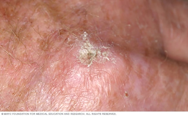
Actinic Keratoses- Skin Cancer But Not Skin Cancer - PeopleBeatingCancer says 9 months ago

[…] When Actinic Keratosis (Pre-Skin Cancer) is Not  […]

Reply
Add Your Reply